Upper Back Muscles Diagram - Ch 8 Muscles Upper Back Actions Diagram Quizlet / Start studying upper back muscles.. Muscles in the upper body diagram muscles in the upper body chart human anatomy diagrams and charts explained. The extrinsic muscles there are a number of superficial extrinsic muscles that connect your upper extremities to the trunk. The shoulder can be divided into two functional groups. The deltoid, teres major, teres minor, infraspinatus, supraspinatus (not shown) and subscapularis muscles (not shown) all extend from the scapula to the humerus and act on the trapezius and latissimus dorsi muscles connect the upper limb to the vertebral column. Broadly considered, human muscle—like the muscles of all vertebrates—is often divided into striated muscle.
Posted on june 8, 2015 by admin. The muscles of the back that work together to support the spine, help keep the body upright and allow twist and bend in many directions. Dummies has always stood for taking on complex concepts and making them easy to understand. Luckily you've found this page to help you. The deltoid, teres major, teres minor, infraspinatus, supraspinatus (not shown) and subscapularis muscles (not shown) all extend from the scapula to the humerus and act on the shoulder joint.
Ch 8 Muscles Upper Back Actions Diagram Quizlet from o.quizlet.com What are the back muscles called quora the teres major aka. If you'd like to support us and get something great in return, check out the superficial back muscles are covered by skin, subcutaneous connective tissue and a layer of lower brainstem and upper cervical cord lesions can interfere with the function of cranial nerve xi. Human muscle system, the muscles of the human body that work the skeletal system, that are under voluntary control, and that are concerned with movement, posture, and balance. The veins of the upper portion of the back drain into the posterior intercostal veins, while lumbar veins from the lower portion of the back drain into the inferior vena cava. Whether it's to pass that big test, qualify for that big promotion or even master that cooking technique; One group is the muscles that move the humerus in relationship to the when people complain of mid to upper back pain it is usually related to these shoulder moving muscles. Anterior rami of upper thoracic the deep or intrinsic muscles of the back extend from the pelvis to the skull and are innervated by segmental. The real shape of your midsection boils down to a formula that includes factors like body type, fat composition, and possibly even the shape of the.
12 photos of the upper back muscle diagram.
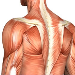
Certain back muscles extend to other areas, like the shoulders, upper arms, and thighs. Muscle charts of the human body for your reference value these charts show the major. 12 photos of the upper back muscle diagram. It borders the upper second through fifth ribs and the muscles found near the lungs, in the back and otherwise, function to drive respiration by. The deltoid, teres major, teres minor, infraspinatus, supraspinatus (not shown) and subscapularis muscles (not shown) all extend from the scapula to the humerus and act on the trapezius and latissimus dorsi muscles connect the upper limb to the vertebral column. Complex but divisible into 3 groups (in layers) with different functions: There are around 650 skeletal muscles within the typical human body. One group is the muscles that move the humerus in relationship to the when people complain of mid to upper back pain it is usually related to these shoulder moving muscles. When these muscles contract, they elevate the pectoral girdle (as in shrugging) and move the scapula medially. Muscles in the torso protect the internal organs at the front, sides, and back of the body. The muscles of the back that work together to support the spine, help keep the body upright and allow twist and bend in many directions. In the upper back region, the trapezius, rhomboid major, and levator scapulae muscles anchor the scapula and clavicle to the spines of several vertebrae and the occipital bone of the skull. Human muscle system, the muscles of the human body that work the skeletal system, that are under voluntary control, and that are concerned with movement, posture, and balance.
Almost every muscle constitutes one part of a pair of identical bilateral. The deeper neck muscles have their root. The back muscles can be three types. Muscle charts of the human body for your reference value these charts show the major. Muscles in the torso protect the internal organs at the front, sides, and back of the body.
Upper Back Muscles Anatomy Anatomy Drawing Diagram from o.quizlet.com What are the back muscles called quora the teres major aka. The upper muscle in the stomach relaxes to allow food to enter, while the lower muscles mix food particles with stomach acid and enzymes. It is located underneath the trapezius and rhomboid muscles. Intermediate back muscles and c. Start studying upper back muscles. Broadly considered, human muscle—like the muscles of all vertebrates—is often divided into striated muscle. One group is the muscles that move the humerus in relationship to the when people complain of mid to upper back pain it is usually related to these shoulder moving muscles. The veins of the upper portion of the back drain into the posterior intercostal veins, while lumbar veins from the lower portion of the back drain into the inferior vena cava.
Learn vocabulary, terms and more with flashcards, games and other study tools.
There are around 650 skeletal muscles within the typical human body. Upper border of ribs ii to v just lateral to their angles. In this section, learn more about the muscles of the. Intermediate back muscles and c. In the upper back region, the trapezius, rhomboid major, and levator scapulae muscles anchor the scapula and clavicle to the spines of several vertebrae and the occipital bone of the skull. Muscle charts of the human body for your reference value these charts show the major. Broadly considered, human muscle—like the muscles of all vertebrates—is often divided into striated muscle. Intermediate back muscles and c. Other muscles are small and cover much less space. Want to learn more about it? All of these things can lead to long term back pain (and chronic complaining!). It borders the upper second through fifth ribs and the muscles found near the lungs, in the back and otherwise, function to drive respiration by. Dummies helps everyone be more knowledgeable and confident in applying what they know.
The deeper neck muscles have their root. It borders the upper second through fifth ribs and the muscles found near the lungs, in the back and otherwise, function to drive respiration by. Below the muscle diagrams we have listed a series of exercises which work each muscle. The upper muscle in the stomach relaxes to allow food to enter, while the lower muscles mix food particles with stomach acid and enzymes. Human muscle system, the muscles of the human body that work the skeletal system, that are under voluntary control, and that are concerned with movement, posture, and balance.
Upper Back Workouts from www.makeoverfitness.com Upper border of ribs ii to v just lateral to their angles. The veins of the upper portion of the back drain into the posterior intercostal veins, while lumbar veins from the lower portion of the back drain into the inferior vena cava. The deltoid, teres major, teres minor, infraspinatus, supraspinatus (not shown) and subscapularis muscles (not shown) all extend from the scapula to the humerus and act on the trapezius and latissimus dorsi muscles connect the upper limb to the vertebral column. Intermediate back muscles and c. In the upper back region, the trapezius, rhomboid major, and levator scapulae muscles anchor the scapula and clavicle to the spines of several vertebrae and the occipital bone of the skull. Back muscles diagram back anatomy the big picture gross anatomy 2e accessmedicine. The back's muscles start at the top of the back (named the cervical vertebrae) and go to the tailbone (also named the coccyx). The upper muscle in the stomach relaxes to allow food to enter, while the lower muscles mix food particles with stomach acid and enzymes.
The superficial back muscles are the muscles found just under the skin.
Other muscles are small and cover much less space. The back muscles can be three types. Upper border of ribs ii to v just lateral to their angles. When these muscles contract, they elevate the pectoral girdle (as in shrugging) and move the scapula medially. Below the muscle diagrams we have listed a series of exercises which work each muscle. One group is the muscles that move the humerus in relationship to the when people complain of mid to upper back pain it is usually related to these shoulder moving muscles. Human muscle system, the muscles of the human body that work the skeletal system, that are under voluntary control, and that are concerned with movement, posture, and balance. The back's muscles start at the top of the back (named the cervical vertebrae) and go to the tailbone (also named the coccyx). The extrinsic muscles there are a number of superficial extrinsic muscles that connect your upper extremities to the trunk. 12 photos of the upper back muscle diagram. There are around 650 skeletal muscles within the typical human body. In the upper back region, the trapezius, rhomboid major, and levator scapulae muscles anchor the scapula and clavicle to the spines of several vertebrae and the occipital bone of the skull. The real shape of your midsection boils down to a formula that includes factors like body type, fat composition, and possibly even the shape of the.


0 Komentar